


The spine, like any structure that performs a support function, is bound to wear out all the time. High static and dynamic loads and local loads from the upper segment are mainly moving cause a decrease in regenerative capacity and gradual degeneration of cartilage and nearby muscle-ligament structures. At the age of 30-35 years, almost everyone shows signs of cervical osteochondrosis to a greater or lesser degree. And while it is impossible to stop the irreversible process of biological aging, it is very possible to slow it down.
Diagnostics
For objective assessment of the condition and detection of degenerative-dystrophic changes in the cervical spine, radiation imaging methods were used:
- normal spondylogography (non-contrast X-ray study in frontal, lateral and oblique projections)
- radiography with functional testing
- MSCT (multislice computed tomography)
- MRI
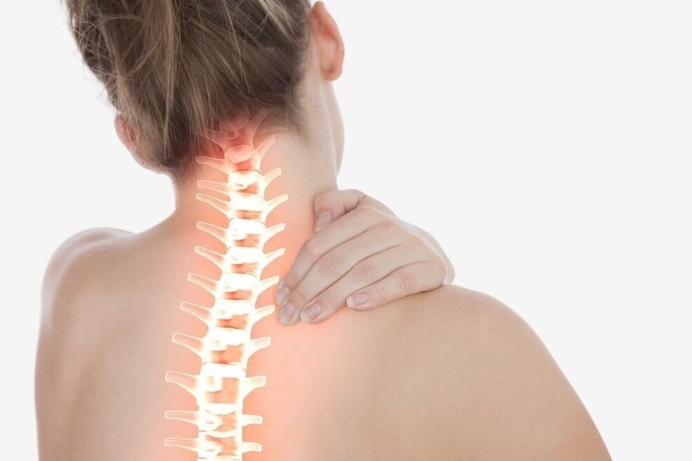
- Spinal survey spondylography is a traditional method of radiological diagnosis of cervical osteochondrosis. With its help, the condition of the vertebral bodies is assessed, the shape, height, degree of deformation and interrelated displacement are determined. On X-ray images, osteophytes, areas of illumination in the focus of bone tissue liquefaction are seen.
- Spondylogography with functional testing is a study aimed at identifying signs of movement disorders. X-rays were performed with constant maximum flexion and elongation of the cervical spine.
- MSCT is a progressive alternative to X-rays. The structures of bones, intervertebral discs, ligament apparatus, spinal canals and spinal cord are seen in more detail on multi -layered photographs.
- Magnetic resonance imaging allows additional visualization of the cartilage and other soft tissue layers of the vertebral joints. The study was prescribed for severe neurological symptoms, to distinguish cervical osteochondrosis from acute intervertebral hernia.
Treatment of cervical osteochondrosis
Treatment of osteochondrosis of the cervical spine aims to relieve pain and slow the progression of pathological processes. It is done in two directions: limiting the impact of unfavorable factors and suppressing the mechanism of disease progression.
Therapeutic and prophylactic measures that reduce the effects of the causative agent include:
- rational selection of work furniture
- use of orthopedic mattresses and pillows
- correction of hearing, vision and posture
- wear special mounting tools
- restrictions on work activities associated with prolonged stay in forced conditions
- adequate physical activity
- proper nutrition
There are many methods of therapeutic correction designed to slow the progression of the degenerative process.
Massage for cervical osteochondrosis
Massage procedures aimed at eliminating inflammation and relieving pain are included in the complex of mandatory therapeutic measures. The most effective types of collar massage:
- classic
- medical (manual)
- point (acupuncture)
- vacuum (canned)
- hardware
Thanks to massage techniques, local blood and lymph circulation is improved, tissue trophism is accelerated, muscle clamps are eliminated, tension from the neck is relieved, muscle tone and elasticity are improved.
Orthopedic collar
To fix the cervical spine in the correct position, special orthopedic devices (Shants collars) are used. Removable structures of various sizes, shapes and degrees of stiffness limit the common pathological position of the head, control movement in the neck, reduce pressure on spinal segments, warm and relax tense muscles and prevent the progression of the disease.
Cervical collars for osteochondrosis are available in several modifications:
Soft flakes made of medical foamor a porous hypoallergenic material having notches to the chin and lower surface of the neck, and retainers. They are used to correct minor disorders in the upper spine, keep the vertebrae in the correct anatomical position and relax the muscles on the shoulder straps.
Pneumatic collar (inflatable)intended for pain prevention, subtle traction and elimination of vertebral artery compression.
Semi -rigid bandageequipped with metal inserts to stably stabilize the intervertebral segment. They significantly limit the distance of movement and contribute to the expansion of the gap between the vertebral bodies.
Rigid corset made of durable plasticdesigned to completely paralyze the cervical spine in a neutral position. Prescribed in the final stages of the disease, accompanied by compression disorders.
Collars for osteochondrosis of the cervical spine are selected by doctors taking into account age, anatomical features and stage of degenerative processes.
Manual therapy
Manual therapy aims to identify and remove blockages in motor segments. The effects of local doses on the vertebral joints help normalize blood flow and blood supply to the brain, eliminate compression (pinching) and restore normal function of nerve fibers. Certain chiropractor manipulations allow you to achieve maximum relaxation, eliminate muscle spasms, cervicogenic headaches arising from damage to the anatomical structure of the neck, and tension headaches.
Acupuncture
Acupuncture, which involves attaching acupuncture needles to bioactive points in the neck and shoulders, is focused on restoring a disturbed energy balance. By stimulating the rapid contraction of sensitive nerve fibers and the release of endorphins and neurotransmitters, acupuncture for cervical osteochondrosis has strong anti-inflammatory and analgesic effects. Thanks to this technique, numbness in the hands, dizziness, tinnitus, increase blood flow and optimize movement.
Physiotherapy
Physiotherapy of degenerative pathology of the spine aims to relieve pain and stimulate the recovery process. The greatest therapeutic effect is provided by:
- UFO
- ultrasound treatment
Frequently asked questions
How to provide relief during acute pain with osteochondrosis of the lumbar spine?
If acute pain is sudden, it is necessary to correct the lower back. This will paralyze the spasmodic muscles and replace the load from them. Then, if possible, lay the patient on his or her back, placing a pillow under the bent knee. To reduce pain, you should take medications with analgesic and anti-inflammatory effects (NSAIDs). Alternatively, you can use an ointment or gel based on diclofenac or its analogues, or apply a cold compress (no more than 10 minutes). It is very important to relieve pressure on the spine and see a doctor as soon as possible.
Is it possible to do physical exercise for lumbar osteochondrosis?
Physical education for lumbar osteochondrosis is not only not prohibited, but also recommended (except for periods of acute pain). However, you should be careful not to let axial loads on the spine and definitely push to squat, jump and lift loads. The training set should be selected by the specialist individually.